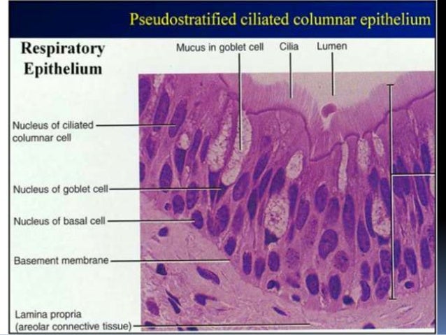
basal lamina basement membrane histology leeds ac uk Connective TissueThe basal lamina lamina layers also known as the basement membrane is a specialised form of extracellular matrix The basal lamina can be organised in three ways 1 it can surround cells for example muscle fibres have a layer of basal lamina around them Connective Tissue Fibres Crossword Extracellular Matrix basal lamina basement membrane D001485Latin membrana basalisTH H2 00 00 0 00005
reference Human AnatomyThe basal lamina is an extracellular matrix that is also known as the basement membrane It is found in many different tissue types and provides support to epithelial cells throughout the body basal lamina basement membrane webster dictionary basement membraneBasement membrane definition is a thin membranous layer of connective tissue that separates a layer of epithelial cells from the underlying lamina propia lamina definition function htmlDirectly beneath the basal lamina sits the reticular lamina portion of the basement membrane which acts as a net of collagen fibers a type of connective tissue that provides support and
is the The basal membrane is the actual plasma membrane on the basal side of an epithelial cell adjacent to the basal lamina basement membrane share improve this answer edited Oct 21 14 at 4 17 basal lamina basement membrane lamina definition function htmlDirectly beneath the basal lamina sits the reticular lamina portion of the basement membrane which acts as a net of collagen fibers a type of connective tissue that provides support and laminaBasal lamina The basal lamina is a fibrous protein matrix that surrounds the neural tube and is also localized to the basal surface of the ectoderm
basal lamina basement membrane Gallery
hcvnl5STmYYqX79qeI7Q, image source: socratic.org
Glomerular_Basement_Membrane_(GBM)_Structure, image source: discovery.lifemapsc.com
picture11335645445832, image source: www.studyblue.com

JCI0629488, image source: www.jci.org
image02727, image source: plasticsurgerykey.com
TUBULES2, image source: www.histology.leeds.ac.uk

histology of trachea and lung 9 638, image source: www.slideshare.net
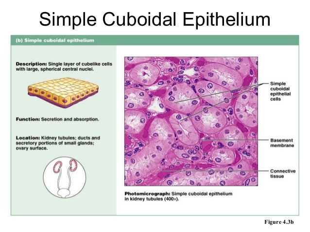
epithelium cellstissues histology 9 638, image source: www.slideshare.net

Cell_adhesion_summary, image source: cellbiology.med.unsw.edu.au
normal_urothelium figureA_Big, image source: www.auanet.org
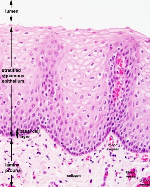
300px Oesophagus_histology_03, image source: embryology.med.unsw.edu.au
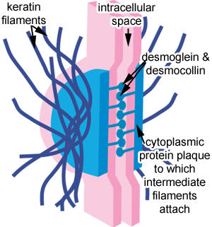
desmosome_diag, image source: histology.leeds.ac.uk

stratified_squamous_31320123308985, image source: www.studyblue.com

1200px Laminin_sketch, image source: en.wikipedia.org

stratified_cuboidal_11320123204834, image source: www.studyblue.com
5 epithelium sp 10 638, image source: www.slideshare.net

Arteries+and+vein+layers, image source: heartsfortheclass.blogspot.com
CDR0000579036, image source: cancerinfo.tri-kobe.org
excretor epitelio transicion espesor, image source: mmegias.webs.uvigo.es
fig30_bd60x6304, image source: www.histology.leeds.ac.uk

Comments