
epithelial basement membrane eyewiki aao Epithelial basement membrane dystrophyEpithelial basement membrane dystrophy EBMD is a disease that affects the anterior cornea causing characteristic slit lamp findings which may result in decreased vision and or recurrent corneal erosions epithelial basement membrane is epithelial basement membrane Fibrogranular deposits below the basement membrane makes the cornea look like a map Dot Fibrogranual deposits above the basement membrane causes the appearance of dots on the cornea Fingerprint Seeing fingerprint lines on the cornea due to the epithelial basement membrane duplicating and regenerating abnormally
dictionary thefreedictionary basement membrane epithelium in animals a layer of covering cells lying on a basement membrane that is called simple epithelium when one cell thick and compound epithelium when several cells thick and usually covers connective tissue embryologically derived from the ECTODERM The cells sometimes have a secretory function and are held together by a cementing substance to form a sheet epithelial basement membrane D001485Latin membrana basalisTH H2 00 00 0 00005 basement membrane Epithelial basement membrane dystrophy EBMD also known as anterior basement membrane disease or map dot fingerprint dystrophy is a common condition that affects the anterior segment of the eye The condition usually affects people over 30 years of age
Basement Membrane Dystrophy EBMD is the most common of the corneal dystrophies Ii is also known as Map Dot Fingerprint Dystrophy and Anterior Basement Membrane Dystrophy ABMD Ii is also known as Map Dot Fingerprint Dystrophy and Anterior Basement Membrane Dystrophy ABMD epithelial basement membrane basement membrane Epithelial basement membrane dystrophy EBMD also known as anterior basement membrane disease or map dot fingerprint dystrophy is a common condition that affects the anterior segment of the eye The condition usually affects people over 30 years of age webeye ophth uiowa edu eyeforum cases 78 EBMD treatment htmEpithelial basement membrane dystrophy fingerprint changes A These changes are easily seen by retroillumination B Duplication of the epithelial basement membrane
epithelial basement membrane Gallery

map dot fingerprint dystrophy 2, image source: webeye.ophth.uiowa.edu

epithelial cell, image source: learnhistology.wordpress.com
c4an00056k f1_hi res, image source: blog.creative-bioarray.com

epithel_comp3, image source: bio.rutgers.edu

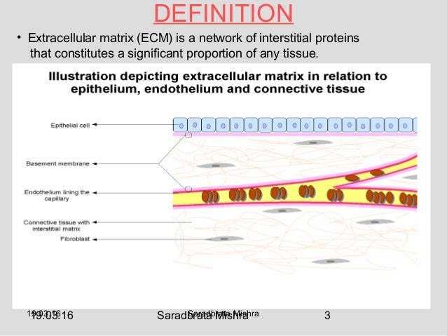
extracellular matrix 3 638, image source: www.slideshare.net
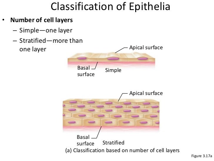
nb ch 3 tissues 4 728, image source: www.slideshare.net
simple_columnar_epithelium13173273552651358909343975, image source: www.studyblue.com

membrane_bilayer, image source: lookfordiagnosis.com


structural_pic2, image source: byjus.com
Zonula+adherens+%28intermediate+junction%29, image source: slideplayer.com


JCI0217302, image source: www.jci.org
c09f008a, image source: pocketdentistry.com

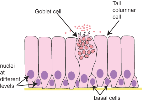
goblet1_001, image source: www.histology.leeds.ac.uk
Simple+Squamous+Epithelium, image source: slideplayer.com

leaky gut syndrome diag, image source: www.top10homeremedies.com

te400t, image source: www.austincc.edu

Toxic_epidermal_necrolysis, image source: smartypance.com


fetal_hemato 14259E7604D40D8811C, image source: www.studyblue.com
Comments