a basement membrane anchors is the basement membrane htmOct 19 2018 The basement membrane membrana basalis is a thin layer of basal lamina and reticular lamina that anchors and supports the epithelium and endothelium Epithelium is a type of tissue that forms glands and lines the inner and outer surfaces of organs and structures throughout the body a basement membrane anchors 25 2016 A basement membrane anchors A muscle tissue to nervous tissue B brain tissue to nervous tissue C connective tissue to muscle tissue D epithelial tissue to connective tissue
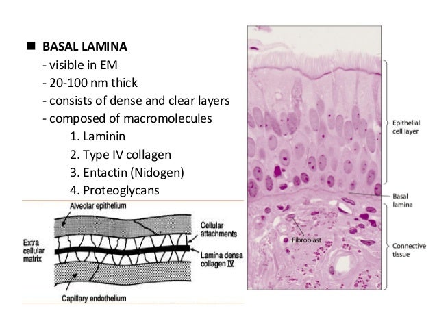
seen with electron microscope the basement membrane is composed of two layers the basal lamina and the underlying layer of reticular connective tissue The underlying connective tissue attaches to the basal lamina with collagen VII anchoring fibrils and fibrillin microfibrils TH H2 00 00 0 00005Latin membrana basalisMeSH D001485Structure Function Clinical significance Further reading a basement membrane anchors ncbi nlm nih gov Journal List HHS Author ManuscriptsCell invasion through basement membrane BM is a specialized cellular behavior critical to many normal developmental events immune surveillance and cancer metastasis A highly dynamic process cell invasion involves a complex interplay between cell intrinsic elements that promote the invasive phenotype and cell cell and cell BM interactions that regulate the timing and targeting of BM transmigration biology fulltext S0962 8924 06 Anchor cell invasion in Caenorhabditis elegans is a simple visually and experimentally accessible model of basement membrane invasion that is beginning to reveal a network of cellular and molecular control mechanisms that regulate the fundamental cellular process of invasion through basement membranes
ap exam tissues flash cards Cutaneous membrane refers to skin Embryonic stem cells growing in a lab dish are bathed in a cocktail of chemicals that cause them to specialize into branching networks of single nucleated cells that pulsate in unison a basement membrane anchors biology fulltext S0962 8924 06 Anchor cell invasion in Caenorhabditis elegans is a simple visually and experimentally accessible model of basement membrane invasion that is beginning to reveal a network of cellular and molecular control mechanisms that regulate the fundamental cellular process of invasion through basement membranes membrane function The basement membrane lies between the epidermis and the dermis keeping the outside layer tightly connected to the inside layer Not even the effects of gravity can destroy this anchoring system
a basement membrane anchors Gallery

picture18 1480D3AECDF34702A14, image source: studyblue.com

image8, image source: www.muhadharaty.com

5 epithelium sp 8 638, image source: www.slideshare.net
figure_04_23_labeled, image source: droualb.faculty.mjc.edu
figure_04_23_labeled 140E4E48D5C5EC9DFC4, image source: www.studyblue.com
4 weeping tiles foundation, image source: nusitegroup.com

TE521_sillplate3_%20PNNL CV_09 16 12, image source: basc.pnnl.gov
da7c25ff9, image source: flylib.com
.jpg?v=2434a81f)
dpc injection cream 6 x 1ltr bba (1200x630 ffffff), image source: www.twistfix.co.uk
brown_20adipose_20tissue_20photo_20with_20label_20copy 143BD7B3E827E55444C, image source: www.studyblue.com
.jpg?v=20b145d4&mode=h)
polyester resin 380ml (gallery), image source: www.twistfix.co.uk

head PBaker Window repair55219 78_0, image source: www.protradecraft.com
.jpg?v=b2c2901d)
brick pin fixing kit (1200x630 ffffff), image source: www.twistfix.co.uk
Simple+Squamous+Epithelium, image source: slideplayer.com

mi201668f1, image source: www.nature.com
.jpg?v=8dbf131f)
brick repair mortar burnt orange (1200x630 ffffff), image source: www.twistfix.co.uk

icf_bracing_scaffolding2, image source: www.quadlock.com

peritoniuem1306455127417, image source: www.studyblue.com
desmosome, image source: antranik.org
Comments