epithelial basement membrane dystrophy eyewiki aao Epithelial basement membrane dystrophyEpithelial basement membrane dystrophy EBMD is a disease that affects the anterior cornea causing characteristic slit lamp findings which may result epithelial basement membrane dystrophy
is epithelial basement membrane Epithelial Basement Membrane Dystrophy EBMD is the most common form of corneal dystrophy It is believed some 2 of the population is affected by it Most patients never develop any symptoms or have some minor discomfort at irregular times epithelial basement membrane dystrophy webeye ophth uiowa edu eyeforum cases 78 EBMD treatment htmEpithelial basement membrane dystrophy fingerprint changes A These changes are easily seen by retroillumination B Duplication of the epithelial basement membrane correlates with the Basement Membrane Dystrophy EBMD is the most common of the corneal dystrophies Ii is also known as Map Dot Fingerprint Dystrophy and Anterior Basement Membrane Dystrophy
basement membrane Epithelial basement membrane dystrophy EBMD also known as anterior basement membrane disease or map dot fingerprint dystrophy is a common condition that affects the anterior segment of the eye The condition usually affects people over 30 years of age epithelial basement membrane dystrophy Basement Membrane Dystrophy EBMD is the most common of the corneal dystrophies Ii is also known as Map Dot Fingerprint Dystrophy and Anterior Basement Membrane Dystrophy eyesofwestwood view article 247 3conxThere is a variety of corneal dystrophies but the two most common are epithelial basement membrane dystrophy EBMD and endothelial cell dystrophy EBMD also known as map dot fingerprint dystrophy is by far the most common of the corneal dystrophies
epithelial basement membrane dystrophy Gallery
map dot fingerprint dystrophy 1, image source: webeye.ophth.uiowa.edu

picture23 14267BD873238BFB78F, image source: www.studyblue.com

Epithelial+%28Anterior%29+Basement+Membrane+Dystrophy+%28EBMD+or+ABMD%29, image source: slideplayer.com
Fig 4A RES, image source: www.eyerounds.org
mapdot corneal dystrophy, image source: webvision.med.utah.edu
Whirlwind 3, image source: eye.keckmedicine.org
meesman 1 LRG, image source: webeye.ophth.uiowa.edu
Fig2, image source: www.reviewofcontactlenses.com
screen_shot_2013 10 14_at_105244_pm 141BAACA0DC5765432F, image source: www.studyblue.com
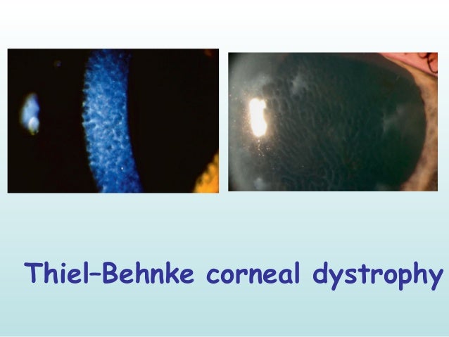
corneal dystrophies 14 638, image source: www.slideshare.net

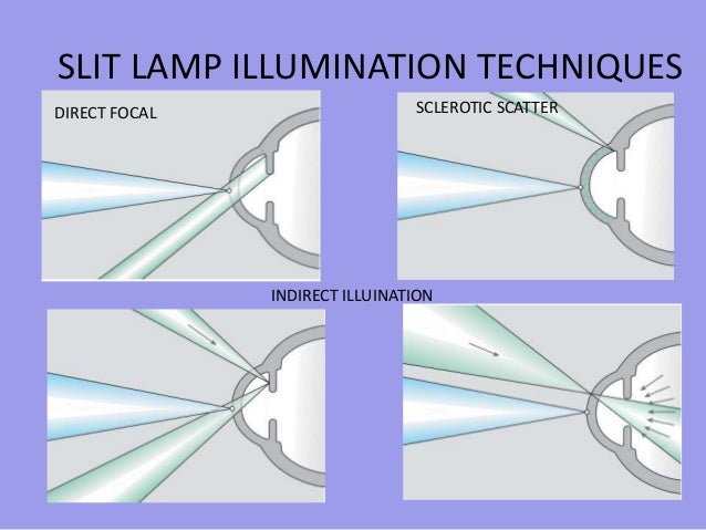
ophthalmology signs around us ii 14 638, image source: www.slideshare.net
picture18 142679F915F4BC4CC96, image source: www.studyblue.com
CLS_June_A10_Fig03, image source: www.clspectrum.com

anatomy and physiology of cornea 22 638, image source: www.slideshare.net

corneal dystrophies 7 638, image source: www.slideshare.net
1704_1716_1, image source: drugster.info
podocyte 11806_2, image source: drugline.org
conjunctivitis_bacterial2_149590, image source: pinterest.com
Epicleritis+vs+Scleritis+ +inflammation+of+episcleral+or+scleral+layers+of+eye, image source: slideplayer.com
Comments